Living organisms are made up of delimited cells by a living barrier, called the plasma membrane, whose structure determines that the organism functions as a harmonious whole. It is precisely here where Dr. Susana Sánchez, a researcher at the Universidad de Concepción, focuses her research, in which she injects fluorescent molecules into living cells and these, through different colors, reveal what their structure is like.
By Monserrat Chávez
/ monsechavez@udec.cl
/ Photographies: Coutesy of FCQ
Fluorescent molecules are tiny particles, much smaller than a cell. They have the ability to absorb light and then emit it, which allows, with a confocal microscope to visualize and to study the small structures that are illuminated.
Thus, when Doctor Susana Sánchez, from the Macromolecular Interactions Laboratory (LIMM) of the Department of Polymers of the Faculty of Chemical Sciences of the Universidad de Concepción, did her doctorate in Fluorescence Spectroscopy, applied to proteins, she realized the great potential of this technique applied to Biomedicine. So, “I did a post-doctorate in the United States in a laboratory that works precisely on that, and there I learned that studies of membrane fluidity using fluorescence spectroscopy in a microscope could help understand abnormalities at the cellular level behind pathologies such as diabetes, Alzheimer’s disease and atherosclerosis.
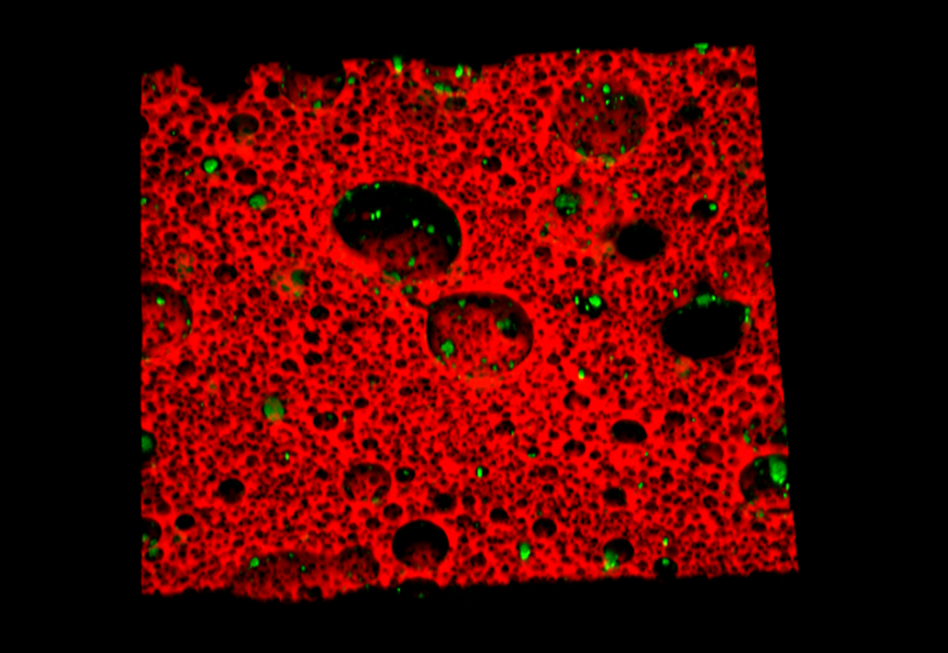
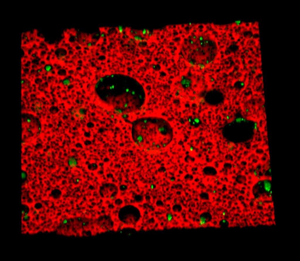
- Fluency image of a neuron
- Polymer with fluorescent nanoparticles
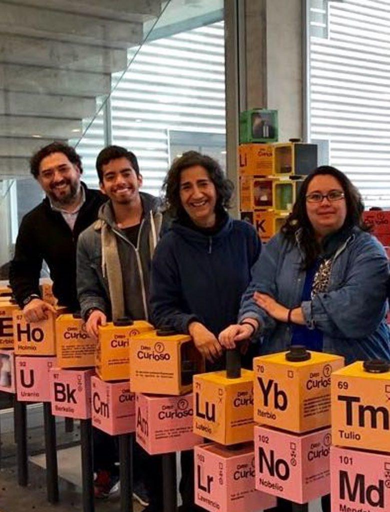
- Macromolecular Interaction Laboratory

For example, the academic says that cardiovascular accidents occur because deposits called atheromas grow in the arteries which can obstruct the passage of blood in small blood vessels. HDL-cholesterol, which appears in blood tests, corresponds to particles responsible for removing cholesterol from the membranes and keeping them with adequate fluidity. In our laboratory we build these particles (rHDL) and use them to study the mechanism of cholesterol extraction and to be able to optimize it”.
In this research, fluorescent molecules act as “reporters” that can be located in specific places within the cell and inform us about what is happening in that place by changing its chemical and physical properties. One of these molecules, called Laurdan, is located in the membranes and changes color depending on whether it is in a compact or fluid environment, that is, it can measure changes in fluidity. “Interpreting the information delivered by fluorescent reporter molecules when they are in a certain place is the line of research of our group”, explains the researcher.
INTERDISCIPLINARY WORK
“Interest in our research may come from different angles; On the one hand, there are people who will be interested in the scientific answers born out of the group’s specific research, while other researchers may be interested in using the research tools we employ, such as microscopy, fluorescence, and macromolecule biochemistry, to be adapted to their own biological and non-biological systems”, explains the expert. “In fact, we have used fluorescence microscopy and associated techniques in collaborations with several researchers from other disciplines in cells and also in polymeric materials, from the Universidad de Concepción. Interdisciplinary support is essential to move forward in science,” she says.
In this sense, “making our work known to the rest of the scientists is very important and in this context, our research has been recently published in the journal Accounts of Chemical Research (ACR), a journal with a multidisciplinary audience and high circulation”, pointed out Doctor Sánchez.
For more information, please contact: susanchez@udec.cl
Last modified: 29 de agosto de 2025